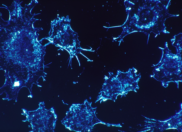
The DNA behaviour of cancer cells has been broken down by scientists using 3D models in a revolutionary new study that could serve as a breakthrough in the treatment of the notorious disease.
The study which validates the employment of 3D samples in the advancement of cancer research and provides insights into new treatments was published in the journal Epigenetics
A first in the field
In what is a first for science, a research team led by Dr. Manel Esteller, Director of the Josep Carreras Leukaemia Research Institute (IJC), demonstrated how 3D models (known as organoids) can now be used to develop a characterization of the DNA make-up - or the epigenetic fingerprint - of human cancer.
Dr Esteller, who is also Chairman of Genetics at the University of Barcelona, explains: "Frequently, promising cancer therapies fail when applied to patients in the real clinical setting. This occurs despite many of these new treatments demonstrating promising results at the preclinical stage in the lab. One explanation is that many of the tumor models used in early research phases are established cell lines that have been growing for many decades and in two dimension (2D) culture flasks. These cancer cells might not completely resemble the features of real tumors from patients that expand into three dimensions (3D)."

"Very recently, it has been possible to grow cancers in the laboratories but respecting the 3D structure: these models are called 'organoids'. We know very little about these cells and if they actually mimic the conformation of the tumor within the body, particularly the chemical behaviors (known as modifications) of DNA that are called epigenetics ("beyond the genetics"), such as DNA methylation.
"What our article solves is this unmet biomedical need in the cancer research field: the characterization of the epigenetic fingerprint of human cancer organoids. The developed study shows that these tumor models can be very useful for the biomedical research community and the pharmaceutical companies developing anti-cancer drugs."
Several useful findings
Specifically looking at 25 human cancer organoids, made available from the American Type Culture Collection (ATTC), Dr. Esteller, an ICREA Research Professor, states that during their research the team made some interesting findings around the properties of the cancer cells.
"First, we found that every cancer organoid retains the properties of the tissue of origin, so this shows that if the samples were obtained from the surgery of a colon or pancreatic cancer, the organoid closely resembles the original primary tumor.
"Second, we discovered that there is no contamination of normal cells, thus, the malignant pure transformed cells can be analyzed without interferences. And finally, the 3D organoid cancers are closer to the patient tumors than the commonly used 2D cell lines."
Enabling further data mining
The study will now be used to help form Big Data, as the team's samples will be shared in easily accessible public databases between researchers to promote more collaborative studies. "This will enable further data mining to produce new cancer discoveries using different biometric approaches or focusing on particular genes," explains Dr. Esteller.
"And most importantly, the characterized cancer organoids can be readily obtainable from a reliable provider (the ATCC) researchers around the world can use the epigenetic information of these sharable samples to develop their own investigations."
(With inputs from agencies)


