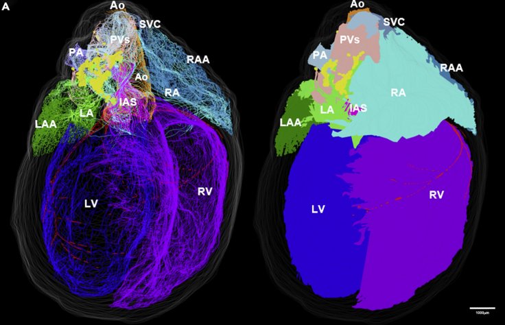
There are neurons in the heart, not only in the brain. Yes, you heard it right. Scientists have now mapped these neurons surrounding the heart for the first time by a 3D heart model. This detailed virtual 3D model that captures the heart's neurons with precision was created by a team led by pathologists James Schwaber and Raj Vadigepalli.
The main role of the heart - as we know - is in circulating oxygen and blood to the whole body, controlled by the brain via the network of nerves between the brain and the heart. But, there are heart's own neurons that surround it, acting as the heart's mini-brain that's known to monitor local heart health. However, its working is not fully known.
Researchers at Thomas Jefferson University, Philadelphia, used the rat's heart as a model and mapped the cardiac nervous system, down to the cellular level. After this, a layer of gene expression data was added, thus creating a 3D heart model. This allows any researchers to use this model in studying the various functions of the heart's neurons with much more precision.
Cutting-Edge Technology

The research paper is published in the Cell journal's iScience. The researchers used knife-edge scanning microscopy, cutting very thin layers of the heart to scan every layer. They used the software, 3Scan to process the information. A diamond knife was used to make fine slices through the heart. Further, they used laser capture microdissection, taking microscope images of each slice, reaching cellular level. Thus, they mapped individual neurons in their positions on the heart.
The scientists used a program called TissueMaker to construct the 3D heart model by manually working out the mapping and contouring the image. By layering gene expression data on each neuron, information about the functions of the cell stored in our DNA can be found. One can measure concentrations of different mRNAs in cells and extract detailed information about the role and functions of cells
Even Doctors Don't Know This

"Many cardiologists aren't even aware there are neurons in the heart, let alone that they are critical to heart health," Schwaber told Freethink. This initiative came as a part of Stimulating Peripheral Activity to Relieve Conditions (SPARC) program where they post their work online for access to any researcher.
Uses
Schwaber said, using the model, one can study the connections, placement, and properties of the heart's neurons. In doing so, they discovered a cluster of neurons that surrounds a "sinoatrial" node in the base of the heart, playing a key role in setting heart rate.
They also found differences between male and female hearts helping scientists know why heart disease differs between the sexes. This was found using rats' hearts, but the team looks at replicating the model with healthy and diseased human hearts.


