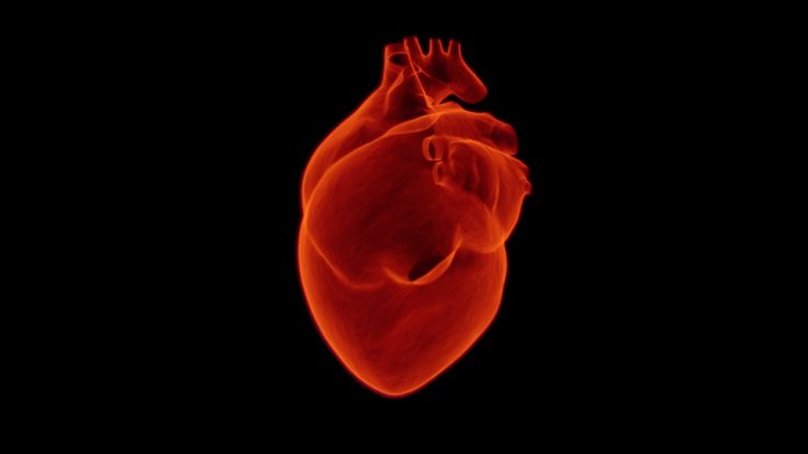
Researchers from the University of Minnesota have managed to create 3D-printed lifelike models of the heart's aortic valve along with the accompanying structures that replicate the accurate feel and look of a real patient.
Michael McAlpine, the senior author of the study, explained, "Our goal with these 3D-printed models is to reduce medical risks and complications by providing patient-specific tools to help doctors understand the exact anatomical structure and mechanical properties of the specific patient's heart."
Improving Outcomes in Patients

These patient-specific organ models, which include 3D-printed soft sensor arrays integrated into the structure, are fabricated using specialized inks and a customized 3D printing process. Such models can be used in preparation for minimally invasive procedures to improve outcomes in thousands of patients worldwide.
The researchers, with the help of medical device company Medtronic, 3D printed what is called the aortic root, the section of the aorta closest to and attached to the heart. The aortic root consists of the aortic valve and the openings for the coronary arteries. The aortic valve has three flaps, called leaflets, surrounded by a fibrous ring.
The model also included part of the left ventricle muscle and the ascending aorta. "Physicians can test and try valve implants before the actual procedure. The models can also help patients better understand their own anatomy and the procedure itself," added McAlpine.
Enabling Doctors to Prepare for Procedures
This organ model was specifically designed to help doctors prepare for a procedure called a Transcatheter Aortic Valve Replacement (TAVR) in which a new valve is placed inside the patient's native aortic valve.
The procedure is used to treat a condition called aortic stenosis that occurs when the heart's aortic valve narrows and prevents the valve from opening fully, which reduces or blocks blood flow from the heart into the main artery. The aortic root models are made by using CT scans of the patient to match the exact shape.
They are then 3D printed using specialized silicone-based inks that mechanically match the feel of real heart tissue the researchers obtained from the University of Minnesota's Visible Heart Laboratories. Commercial printers currently on the market can 3D print the shape, but use inks that are often too rigid to match the softness of real heart tissue.
Improvement and Integration
On the flip side, the specialized 3D printers at the University of Minnesota were able to mimic both the soft tissue components of the model, as well as the hard calcification on the valve flaps by printing an ink similar to spackling paste used in construction to repair drywall and plaster.
Physicians can use the models to determine the size and placement of the valve device during the procedure, said the study published in Science Advances, a peer-reviewed scientific journal. "As our 3D-printing techniques continue to improve and we discover new ways to integrate electronics to mimic organ function, the models themselves may be used as artificial replacement organs," McAlpine said.
(With inputs from agencies)


